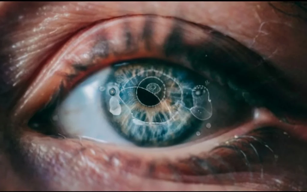
Ophthalmic Anatomy: A Focus on Vision for Specialized Biology Assignments

In the intricate world of biology, one facet that captivates researchers and students alike is the mesmerizing realm of ophthalmic anatomy. As we delve into the complex web of structures that comprise the human eye, a profound understanding of vision emerges, making it a captivating subject for specialized biology assignments. The eye, often referred to as the window to the soul, is a marvel of evolutionary engineering, equipped with an array of specialized components that work in harmony to perceive the visual stimuli that surround us. This blog will serve as a comprehensive guide for those undertaking specialized biology assignments, offering a focused exploration into the intricacies of ophthalmic anatomy. If you need assistance with your anatomy assignment, delving into the complexities of ophthalmic anatomy provides an intriguing avenue for exploring the intricacies of biological structures and their functions, enhancing your understanding of specialized anatomical systems.
At the core of this exploration lies the intricate architecture of the eye, where various components such as the cornea, lens, retina, and optic nerve play crucial roles in the visual process. Understanding how these elements interact enables a deeper comprehension of vision and the physiological processes that underlie it. Specialized assignments in ophthalmic anatomy provide students with the opportunity to dissect these components, both metaphorically and, in some cases, literally, enhancing their grasp of the intricacies involved. Moreover, the blog will shed light on the fascinating adaptations that have occurred throughout the evolutionary journey, shaping the eye into the sophisticated organ we rely on daily.

The blog will also delve into the pathophysiology of vision-related disorders, offering insights into conditions such as myopia, hyperopia, astigmatism, and more. By examining the anatomical aberrations associated with these disorders, students can develop a holistic understanding of the challenges individuals face in maintaining optimal vision. Furthermore, the integration of cutting-edge research and technological advancements in ophthalmology will be highlighted, showcasing the dynamic nature of this field and its relevance to contemporary healthcare.
In addition to the biological aspects, the blog will emphasize the interdisciplinary nature of ophthalmic anatomy, touching upon its connections with neuroscience, genetics, and even psychology. Vision is not merely a biological phenomenon; it is a sensory experience that intertwines with various facets of human existence. As students navigate through their specialized assignments, they will be encouraged to consider the broader implications of ophthalmic anatomy, fostering a holistic perspective that transcends traditional disciplinary boundaries. In essence, this blog serves as a guiding beacon, illuminating the intricate tapestry of ophthalmic anatomy and encouraging students to explore the limitless possibilities within this captivating realm of biology.
The Intricacies of Ophthalmic Anatomy
The intricacies of ophthalmic anatomy unfold a captivating narrative of the marvelously complex structures that constitute the human eye. At the heart of this anatomical spectacle lies a symphony of specialized components, each playing a pivotal role in the extraordinary process of vision. The cornea, a transparent outer layer, acts as the eye's protective shield and refracts incoming light. Adjacent to it, the crystalline lens further refines and focuses the light onto the retina, a light-sensitive layer lining the back of the eye that converts visual stimuli into nerve signals. The optic nerve then carries these signals to the brain, where the intricate dance of neural processing transforms them into the rich tapestry of our visual experiences.
Exploring ophthalmic anatomy unveils the sheer elegance of evolutionary design. The eye has undergone millennia of refinement, adapting to diverse environmental challenges to become the sophisticated organ we rely on daily. From the basic photoreceptor cells in the retina, known as rods and cones, to the intricacies of the ciliary muscles responsible for lens accommodation, every facet of the eye reflects a fascinating evolutionary journey.
Beyond the surface-level examination of anatomical structures, a comprehensive understanding of ophthalmic anatomy also delves into the pathophysiology of vision-related disorders. Myopia, hyperopia, astigmatism, and other conditions are dissected to reveal the minute deviations from the norm that can profoundly impact visual acuity. This exploration enables a deeper grasp of the challenges individuals face in maintaining optimal vision and opens avenues for innovative solutions in the fields of optometry and ophthalmology.
Moreover, the interdisciplinary nature of ophthalmic anatomy emerges as a central theme. Connections with neuroscience, genetics, and psychology underscore the holistic approach required to comprehend the full spectrum of vision. The eye is not merely a biological entity but a sensory gateway that intertwines with various dimensions of human existence. As we unravel the intricacies of ophthalmic anatomy, we are prompted to consider the broader implications and applications in fields ranging from medical research to advancements in visual technology.
Navigating the Lens: A Closer Look at Accommodation
Navigating the Lens: A Closer Look at Accommodation invites us to embark on an enlightening exploration of the intricate mechanisms governing vision, focusing specifically on the remarkable phenomenon of accommodation. This physiological process, orchestrated by the crystalline lens within the eye, plays a pivotal role in our ability to shift our focus seamlessly between objects at varying distances, ensuring clarity and precision in our visual experiences.
At the heart of accommodation lies the dynamic nature of the crystalline lens, a transparent structure positioned behind the iris. This lens is not a static entity but rather a marvel of adaptability, adjusting its shape to alter the refractive power of the eye. This shape-shifting ability is crucial for maintaining sharp vision across different distances, a feat achieved through the coordinated action of ciliary muscles surrounding the lens.
When the eye is focused on a nearby object, the ciliary muscles contract, causing the lens to thicken and increase its curvature. This increased curvature enhances the refractive power of the lens, allowing the eye to converge light rays onto the retina, thus creating a clear image of the close-up object. Conversely, when the gaze shifts to a distant target, the ciliary muscles relax, and the lens becomes flatter, reducing its refractive power. This adjustment ensures that parallel light rays coming from distant objects are focused precisely onto the retina, maintaining visual acuity.
The blog will also delve into the intricacies of presbyopia, a common age-related condition affecting accommodation. As we age, the crystalline lens gradually loses its flexibility, making it more challenging for the eye to adjust focus for close-up tasks. Understanding the biomechanics of accommodation provides valuable insights into the etiology of presbyopia, offering a foundation for developing corrective measures such as reading glasses or multifocal lenses.
Moreover, the blog will shed light on the influence of external factors on accommodation, including lighting conditions, visual habits, and technological advancements. The pervasive use of digital devices, for instance, has prompted discussions on the potential impact on accommodative function and the importance of incorporating visual breaks into our daily routines.
The Retina and the Miracle of Photoreception
The retina, a delicate and intricate layer lining the back of the eye, stands as a testament to the miracle of photoreception, the process through which light is converted into electrical signals, laying the foundation for vision. As the primary sensory organ for sight, the retina orchestrates a symphony of specialized cells, most notably rods and cones, to capture and interpret the visual stimuli that surround us.
Rods and cones, the two primary types of photoreceptor cells embedded in the retina, play distinctive roles in shaping our visual experiences. Rods, more abundant and sensitive to low levels of light, are crucial for vision in dimly lit environments, contributing to our ability to see in low-light conditions. On the other hand, cones, concentrated in the central region of the retina known as the fovea, specialize in daylight vision, color perception, and fine detail. The exquisite arrangement of these photoreceptor cells across the retina reflects an evolutionary precision that optimizes our visual acuity in diverse environmental settings.
The process of photoreception begins with the absorption of light by specialized pigments in rods and cones. This triggers a cascade of biochemical events, leading to the generation of electrical signals. These signals are then transmitted through the intricate network of nerve cells in the retina, ultimately converging at the optic nerve, which serves as the communication highway between the eye and the brain.
The journey of these electrical signals to the brain unveils the marvel of neural processing, where the visual information is decoded, interpreted, and transformed into the rich tapestry of our perceptual experience. The brain's visual cortex, a sophisticated region responsible for processing visual stimuli, collaborates with various structures to construct the vibrant mosaic of colors, shapes, and depths that constitute our visual reality.
The study of the retina and photoreception extends beyond basic anatomy and physiology, delving into the realms of vision-related disorders and groundbreaking research. Disorders such as retinal degeneration underscore the fragility of this intricate system, while ongoing research endeavors explore innovative treatments and technologies to restore or enhance vision.
Decoding Visual Information: The Role of Photoreceptor Cells
Decoding Visual Information: The Role of Photoreceptor Cells invites us to delve into the captivating world of the eye's sensory machinery, focusing on the pivotal players in visual perception—photoreceptor cells. These specialized cells, predominantly rods and cones, form the foundation of the retina and are instrumental in capturing and transducing light into electrical signals that initiate the complex process of vision.
At the forefront of this exploration is the distinction between rods and cones, each endowed with unique characteristics suited to specific visual tasks. Rods, abundant in the peripheral regions of the retina, excel in low-light conditions and are responsible for peripheral vision. Their heightened sensitivity allows us to navigate dimly lit environments, contributing to our overall visual awareness. On the other hand, cones, concentrated in the central region called the fovea, are essential for daylight vision, color discrimination, and fine visual details. This specialization equips us with the ability to discern colors, read, and appreciate intricate visual stimuli.
The blog will unravel the intricacies of the phototransduction process, wherein light energy is converted into electrical signals. Photoreceptor cells contain light-sensitive pigments, rhodopsin in rods and various opsins in cones, which undergo a series of molecular changes upon light exposure. This cascade of events leads to the generation of electrical impulses, forming the basis of the signals transmitted to the brain.
Moreover, the discussion will extend to the concept of visual adaptation, where photoreceptor cells dynamically adjust their sensitivity to changes in ambient light. This mechanism ensures that our eyes can function optimally across a broad range of lighting conditions, from bright sunlight to subdued moonlight, providing a seamless visual experience.
The blog will also highlight the vulnerabilities of photoreceptor cells, emphasizing their susceptibility to degenerative conditions like retinal diseases. Understanding these conditions sheds light on the fragility of our visual system and underscores the importance of ongoing research for therapeutic interventions.
Furthermore, the blog will touch upon emerging technologies and research endeavors aimed at enhancing our understanding of photoreceptor function. From advanced imaging techniques that offer unprecedented views of these cells in action to innovative treatments for vision-related disorders, the intersection of technology and ophthalmic research holds promise for the future of visual healthcare.
Neural Pathways and Vision Processing
The realm of vision processing unveils a captivating narrative of neural pathways, intricate networks that serve as the conduits for visual information from the eyes to the brain, where the magic of perception unfolds. The journey begins with the optic nerve, a bundle of fibers that carries electrical signals generated by the retina to the brain's visual processing centers. This initial leg of the neural pathway marks the critical link between ocular reception and cerebral interpretation, setting the stage for the intricate dance of neural processing.
As the optic nerve extends into the brain, it traverses various regions, including the optic chiasm, where fibers from the nasal halves of each retina cross over. This crossing allows the brain to integrate information from both eyes, creating a cohesive and binocular visual experience. From the optic chiasm, the signals continue along the optic tracts, reaching the lateral geniculate nucleus (LGN) in the thalamus, a vital relay station for sensory information. The LGN serves as a nexus where visual signals undergo preliminary processing before being transmitted to the visual cortex.
The visual cortex, located in the occipital lobes at the back of the brain, is the epicenter of vision processing. Comprising multiple areas, each specializing in distinct aspects of visual perception, the visual cortex orchestrates the intricate decoding and interpretation of the electrical signals received from the eyes. Here, the brain discerns shapes, colors, motion, and spatial relationships, constructing a coherent and nuanced representation of the visual world.
The journey through neural pathways and vision processing is not a one-size-fits-all experience. Parallel processing streams within the visual cortex allow for the simultaneous analysis of different visual features. For instance, the ventral stream, often referred to as the "what" pathway, specializes in object recognition and identification, while the dorsal stream, known as the "where" or "how" pathway, processes information related to motion, spatial relationships, and depth perception.
This intricate dance of neural processing is not limited to the initial stages of visual perception; it extends to higher-order cognitive functions, shaping our visual experiences in complex ways. Emotions, memories, and attention further modulate the neural pathways, influencing what captures our focus and how we interpret visual stimuli.
The Journey to Visual Perception: Exploring Visual Pathways
The Journey to Visual Perception: Exploring Visual Pathways" invites us on an illuminating expedition through the intricate neural circuits that underpin the extraordinary process of visual perception. This exploration begins with the optic nerve, the primary conduit through which visual information travels from the eyes to the brain, and unfolds into the dynamic interplay of visual pathways that shape our perceptual experiences.
At the outset, the blog will spotlight the optic nerve's pivotal role as it carries electrical signals generated by photoreceptor cells in the retina to the brain. The crossing of fibers at the optic chiasm, where information from the nasal halves of each retina converges, allows for the integration of visual input from both eyes, laying the foundation for binocular vision and depth perception.
The journey continues along the optic tracts, traversing through the thalamus, specifically the lateral geniculate nucleus (LGN), which serves as a crucial relay station for visual information. The LGN acts as a gatekeeper, modulating and refining visual signals before dispatching them to the visual cortex in the occipital lobes.
The visual cortex takes center stage in the exploration, comprising distinct areas dedicated to processing various aspects of visual stimuli. The ventral stream, often referred to as the "what" pathway, specializes in object recognition and identification, contributing to our ability to discern shapes and faces. In contrast, the dorsal stream, known as the "where" or "how" pathway, processes information related to motion, spatial relationships, and depth perception, crucial for navigation and interaction with the environment.
The blog will delve into the intricacies of parallel processing within the visual cortex, allowing for the simultaneous analysis of different visual features. This parallelism enables the brain to construct a comprehensive and cohesive representation of the visual world, incorporating aspects like color, form, and motion.
Additionally, the discussion will extend to higher-order cognitive influences on visual perception, such as the role of attention, memory, and emotion in shaping our subjective experiences. The brain's ability to filter, prioritize, and interpret visual stimuli is integral to our capacity to make sense of the complex and dynamic visual environment.
Conclusion:
In conclusion, the exploration of ophthalmic anatomy within the context of specialized biology assignments serves as a captivating journey into the depths of the eye's intricate architecture and the miracle of vision. This focused study not only unravels the anatomical marvels of the cornea, lens, retina, and optic nerve but also delves into the evolutionary tapestry that has shaped the eye into a sophisticated sensory organ. Through specialized assignments, students are afforded the opportunity to dissect, both figuratively and literally, the complexities of ophthalmic anatomy, fostering a profound understanding of the interplay between structure and function.
Moreover, this journey extends beyond mere anatomical exploration, embracing the pathophysiology of vision-related disorders and the interdisciplinary connections with fields such as neuroscience, genetics, and psychology. As students navigate through their assignments, they are encouraged to consider the broader implications of ophthalmic anatomy, recognizing its relevance not only in biological contexts but also in the realms of healthcare, technology, and even the understanding of human experience.
Ultimately, the blog sheds light on the dynamic and evolving nature of ophthalmic anatomy, integrating cutting-edge research and technological advancements. By emphasizing the holistic perspective required to comprehend the intricacies of vision, the blog serves as a guiding beacon for those venturing into the captivating realm of specialized biology assignments, inspiring a profound appreciation for the complexities that underscore the miraculous gift of sight.